The Best Way to Beat Breast Cancer? Early Detection. And You Can Help!
It’s scary to think that 1 in 8 Canadian women will develop breast cancer in her lifetime! However, the good news is that the earlier breast cancer is detected, the better a woman’s chance is of conquering the disease. And you can help.
HGH needs our help to purchase 3D mammography technology, which not only provides a more detailed image, but enables a simple exam that is quicker and less painful. Most importantly, it can also detect cancer even before symptoms appear!
This modern and versatile technology will help throughout the entire breast health continuum, from screening to biopsies to surgery to specimen evaluation.
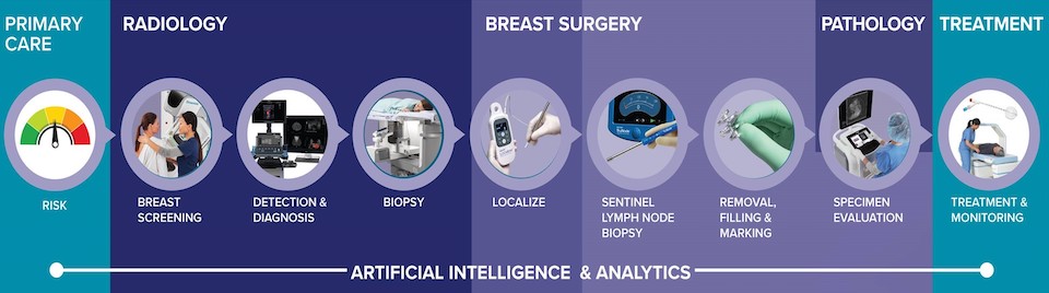
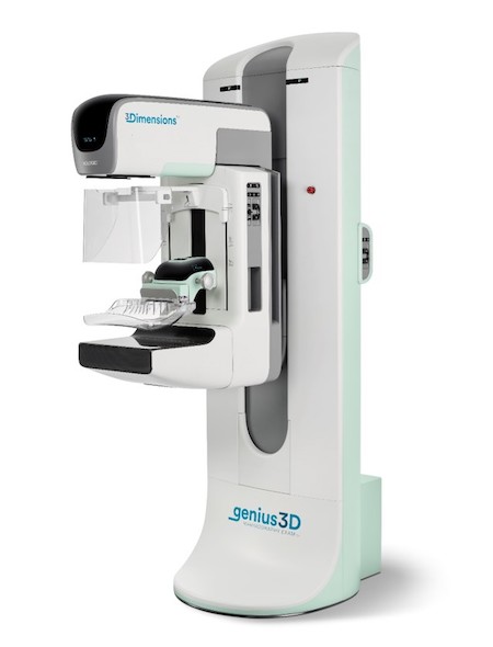
The screening device, called breast tomosynthesis, is an advanced type of breast imaging that uses low-dose X-rays and computer reconstruction to create a 3D image of the breast. The images provide outstanding detail and clarity, enabling the detection of up to 65% more invasive breast cancers than conventional 2D technology!
A new mammography unit would allow HGH to continue offering stereotactic biopsies, which help diagnose cancerous cells in tissue. It would even allow biopsy for small suspicious calcifications that the current unit at HGH does not. The new mammography technology is a vacuum assisted breast biopsy system that integrates tissue acquisition, real-time imaging and verification, as well as post biopsy handling. This removes unnecessary steps to streamline and shorten procedure times by an average of 13 minutes.
To prepare for surgeries, the new mammography system will allow for wire and radioactive free localization of abnormal tissue to allow surgeons to locate even the smallest non-palpable tumours. It is designed to be easy to use and to help surgeons remove lesions more efficiently, while also increasing patient comfort.
The system will also allow better post-surgery sample quality evaluation – right in the surgical suite, making this step a lot more efficient.
As you can see, donating towards this important advanced mammography technology will contribute to significant improvement in the early detection and diagnosis of breast cancer. Your gift will save lives!
Use our secure portal to make an online donation right now.